Retinal conditions we diagnose and treat
The retina is the thin, transparent film that turns light into sight; when it fails, no other eye surgery can help. GS Eye Center's retina clinic in Gangnam, Seoul treats nine retinal conditions with injections, laser and sutureless vitrectomy, performed directly by retina-trained specialists.
When should you see a retina specialist?
Retinal disease can distort vision, and severe disease can end in blindness. Early detection and consistent treatment decide the outcome, so take these seven symptoms seriously.
Sudden drop in vision
Vision that falls quickly, over hours or days, rather than drifting slowly.
Dimness that will not clear
A murky, dim feeling that artificial tears and blinking do not fix.
Flashes of light
Streaks or bursts of light in front of the eye (photopsia).
One side of the view missing
Losing part of the visual field on one side.
A curtain over your vision
A shadow that spreads like a curtain being drawn across the eye.
A sudden swarm of floaters
More dots or fly-like shapes drifting in front of the eye than before.
Bent lines, missing patches
Straight edges look wavy, or objects appear to have holes in them.
Any of these? Get examined.
Some retinal conditions damage vision within hours. Tell us what you are experiencing and we will advise on timing.
Start a free consultationWhat we treat, and how
Each condition below is managed at GS with injections, laser, or vitrectomy surgery, after a work-up on our retinal diagnostic lineup.
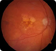 Injections
Macular degeneration
Change in the macula, the part of the retina that receives light most sharply, causes vision loss. Straight lines can look bent (metamorphopsia), and bleeding can block part of the view. Treated with specialist-administered injections.
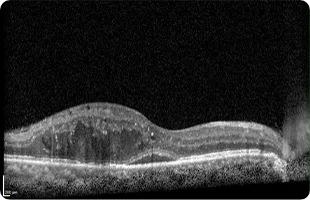 Laser · Injections · Surgery
Diabetic retinopathy
High blood sugar disturbs the fine circulation of the retina, leading to macular edema and vitreous hemorrhage. Early stages often have no symptoms, so regular precision screening matters if you have diabetes.
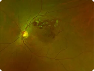 Laser · Injections
Retinal vein occlusion
A clot blocks a retinal vein, which can rupture and bleed into the retina. Small occlusions may only show up on screening; blockage of a large vein can reduce vision within hours and needs prompt care.
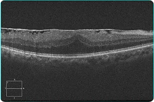 Surgery (vitrectomy)
Epiretinal membrane
A semi-translucent membrane forms on the retinal surface and disturbs macular function; as it thickens it causes swelling and wrinkling. Vision loss and distortion usually progress slowly. Removing the membrane can partially improve vision over several months.
 Surgery + gas
Macular hole
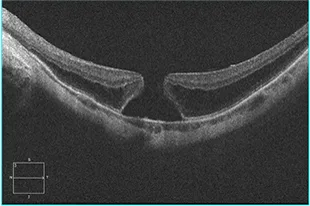
A small hole forms in the macula, mostly with age. Onset can be sudden and progression fast; untreated it can reach blindness. Causes blur, distortion and a central blind spot. Treated by vitrectomy with membrane peeling and a gas bubble, followed by face-down recovery.
 Surgery (vitrectomy)
Retinal detachment
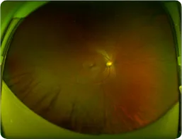
The retina separates from the wall of the eye. Vessels damaged by hypertension or diabetes can end in tractional detachment, which carries a poor outlook once advanced. A spreading shadow or curtain calls for urgent examination.
 Laser · Surgery
Dislocated intraocular lens
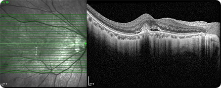
A lens implanted during cataract surgery slips out of position, often from eye rubbing, impact, or inflammation. It causes cloudy vision, double images and glare, and can damage tissue, raise eye pressure and inflame the eye.
 Injections
Myopic choroidal neovascularization
In high myopia, abnormal new vessels grow beneath the macula, causing vision loss and distortion; bleeding can block part of the view. Treated with injection therapy.
 Laser · Injections
Central serous chorioretinopathy
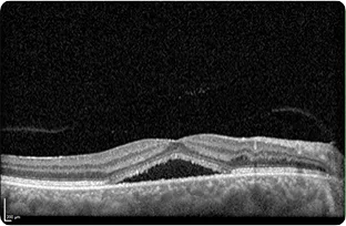
Fluid collects under the central macula and lifts a small area of retina. Vision loss depends on how much fluid there is and how far it spreads; distortion and a central dark spot are common.
How macular disease changes what you see
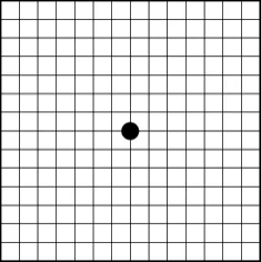
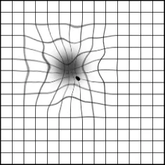
Several of these conditions, macular degeneration among them, announce themselves as distortion: straight lines bend, and a dark or empty patch settles near the center of vision. The grids show the difference between a healthy macula and an affected one.
If a grid ever looks like the right-hand image to you, book a retinal examination rather than waiting for it to pass.
Condition, symptoms, treatment
| Condition | Common symptoms | Treatment at GS |
|---|---|---|
| Macular degeneration | Vision loss, bent lines, blocked patch if bleeding | Injections |
| Diabetic retinopathy | Often none early; macular edema, vitreous hemorrhage later | Laser, injections, vitrectomy |
| Retinal vein occlusion | None (small veins) to vision loss within hours (large veins) | Laser, injections |
| Epiretinal membrane | Slowly progressing vision loss and distortion | Vitrectomy |
| Macular hole | Sudden vision loss, blur, distortion, central blind spot | Vitrectomy + membrane peel + gas |
| Retinal detachment | Curtain-like shadow, field loss | Vitrectomy (gas or silicone oil) |
| Dislocated intraocular lens | Cloudy vision, double images, glare, pressure rise | Laser, vitrectomy |
| Myopic choroidal neovascularization | Vision loss, distortion, blocked patch if bleeding | Injections |
| Central serous chorioretinopathy | Vision loss, distortion, central dark spot | Laser, injections |
Questions patients ask about retinal disease
Age-related macular degeneration is a major cause of blindness worldwide, and the most common cause among people over 60 in Western countries. The dry form rarely leads to blindness; over 90% of AMD blindness comes from the wet form, which, left untreated, can lead to blindness within two years. Early diagnosis is the deciding factor. With severe myopia on the rise, the age of onset is also falling.
Yes. Early diabetic retinopathy typically has no symptoms at all. After 20 years of diabetes, retinopathy is reported in 99% of type 1 and 60% of type 2 patients, and once it develops, controlling blood sugar alone rarely reverses it. Untreated, it can progress from the non-proliferative to the proliferative stage, with new vessels, bleeding inside the retina, and retinal detachment threatening sight.
A sudden drop in vision, a curtain-like shadow, flashes of light, a sudden swarm of floaters, or losing one side of your visual field. Blockage of a large retinal vein can reduce vision within hours, so do not wait these out. Contact us and describe what you are experiencing.
The retina clinic is in the hands of Dr. Park Sungwook and Dr. Lee Jiyoung, both trained in retina at university hospitals. See why patients choose GS for retinal care.
Vitrectomy at GS uses a 25-gauge (0.5 mm) sutureless technique, so admission, surgery and discharge happen on the same day. Macular hole and retinal detachment cases that receive a gas or silicone-oil fill need face-down positioning and about two to three weeks of reduced vision afterward. Details are on the treatments page.