The exam decides the surgery. Not the other way around.
Before any procedure is scheduled, GS Eye Center screens each patient on a diagnostic lineup of more than 20 instruments across six categories: corneal analysis, retinal imaging, refraction, dry-eye testing, ocular biometry and visual field. The examination visit takes about two hours, and its results determine which procedure — if any — is offered.
How is my cornea measured?
Seven instruments cover corneal shape, thickness, cell health and surface condition — the numbers that decide whether laser correction is safe for you.
| Instrument | What it measures |
|---|---|
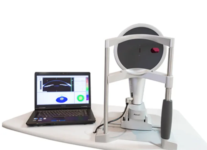
| PENTACAM (corneal topography) | Tomographic imaging of each corneal layer down to 0.002 mm cell-level detail; automatically analyzes endothelial cell count, shape, size and density |
| GALILEI G4 (corneal topography) | Combines anterior segment CT with topography for a 3D analysis; in one second it captures anterior and posterior curvature, anterior chamber depth, corneal diameter and central thickness |
| K5M-TOPO (corneal topography) | Renders corneal curvature and topography maps; the clinic classes it as essential for cataract surgery and Ortho-K (Dream Lens) fitting |
| SPECULAR (endothelium) | Counts and measures corneal endothelial cells — the transparency-related layer — including post-surgical cell loss |
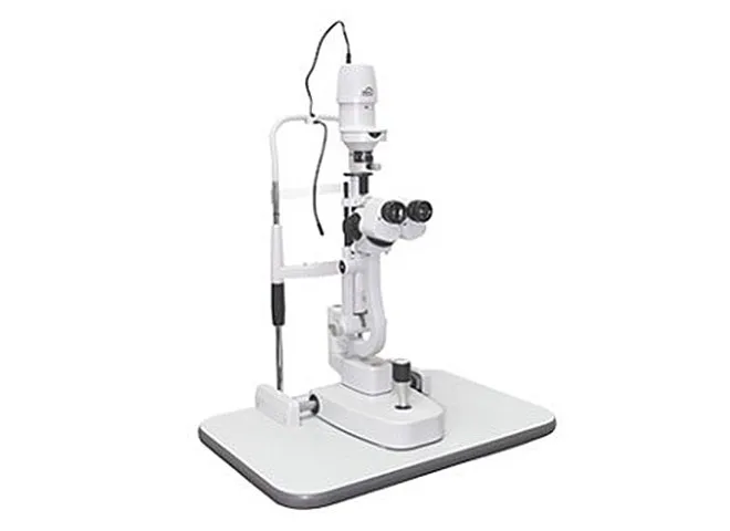
| SLIT LAMP (microscope) | Up to 40x magnification; the doctor directly inspects the conjunctiva, cornea, anterior chamber, iris, lens and front of the vitreous |
| IOP / PACHY | Intraocular pressure and congenital corneal thickness; poorly controlled pressure can damage the optic nerve or relate to conditions like keratoconus |
| FLUORESCEIN (staining) | Dyes the tear film to reveal the presence and extent of corneal surface damage |
What checks the back of my eye?
Six imaging systems examine the retina, optic nerve and the blood vessels behind them — screening that also protects vision-correction candidates from hidden retinal problems.
| Instrument | What it does |
|---|---|
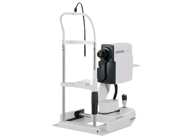
| Spectralis OCT | Images retinal and choroidal blood vessels without contrast dye, tracking the moving retina to catch fine changes in lesions; supports diagnosis of glaucoma, macular degeneration, diabetic retinopathy and retinal vein occlusion |
| FUNDUS (non-mydriatic camera) | Photographs a 45-degree field of the retina, macula and optic disc without dilating drops |
| OPTOS Daytona | Ultra-widefield imaging of about 200 degrees — roughly 80% of the retina — without dilation, reaching the retinal periphery quickly |
| A-SCAN & B-SCAN (ultrasound) | Measures structures inside the eye by ultrasound and reveals vitreous hemorrhage or retinal detachment; A-scan helps when a dense cataract blocks optical measurement |
| Cirrus HD OCT | Rapid high-resolution 3D imaging of retinal structure; its nerve-fiber-layer analysis can detect early glaucoma damage |
| Spectralis HRA | Runs fluorescein (FAG) and ICG angiography at the same time — retinal and choroidal vessel studies in one sitting |
How is my prescription verified?
Four methods are cross-checked rather than trusting a single machine reading.
| Test | How it works |
|---|---|
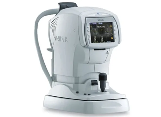
| ARK (auto refraction) | The baseline instrument: you watch a hot-air-balloon target while it measures refractive power, corneal curvature, pupil size and visible iris diameter |
| MR (manifest refraction) | An examiner changes trial lenses while asking what you see — uncorrected vision, refractive error and corrected vision measured in your eye's everyday state |
| CR (cycloplegic refraction) | Drops temporarily paralyze the eye's focusing muscle, then refraction is repeated; comparing MR and CR gives a more accurate prescription |
| Skiascopy / Retinoscopy | A light beam is shone into the eye and the reflex movement read directly — an objective measurement that does not depend on your answers |

Why is dry eye tested before surgery?
Tear quality affects both candidacy and recovery, so it gets its own battery of four tests.
| Test | What it shows |
|---|---|
| K5M | Measures tear-film depth and break-up time, and images the meibomian glands — an overall dry-eye workup |
| Schirmer's test | A paper strip on the lower lid measures tear production over a set time; 10–15 mm is the normal range |
| Fluorescein staining | Dyes the tear layer so its condition can be observed directly |
| Slit lamp | Up to 40x magnification to check for corneal scratches and inspect the tear layer |
How is the eye itself measured?
Biometry drives lens-power calculation for cataract and lens-implant surgery, and confirms corneal thickness for laser candidacy.
| Instrument | What it measures |
|---|---|
| A-SCAN & B-SCAN | Ultrasound measurement of lengths inside the eye, effective even when a dense cataract blocks optical instruments |
| AL-SCAN (optical biometer) | Uses infrared light to rapidly measure axial length, anterior chamber depth, corneal thickness, corneal power, pupil size and visible iris diameter for intraocular lens calculation |
| PACHYMETRY (non-contact) | Corneal thickness — the number that can rule LASIK or LASEK in or out — measured quickly without touching the eye |
| IOL MASTER 700 | Non-contact laser biometry for intraocular lens power, faster and more precise than ultrasound methods; unlike the previous IOL Master 500, it also measures posterior corneal astigmatism |
What about my optic nerve?
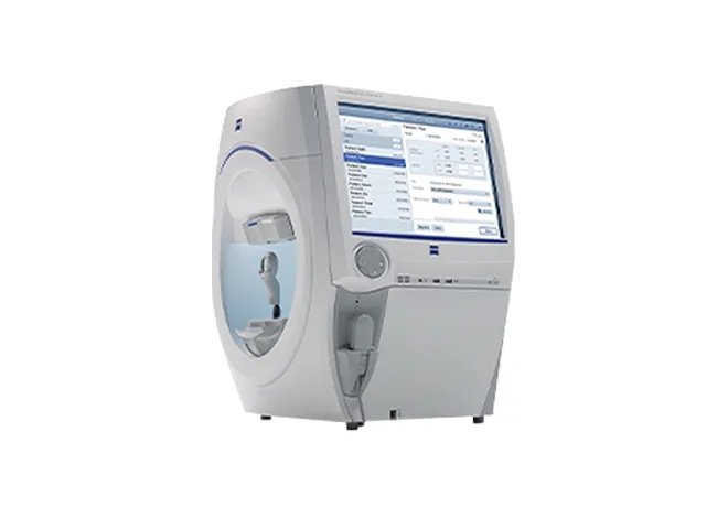
Mapping optic nerve damage
With your chin fixed in place, you press a handheld button each time you notice a light — mapping the sensitivity of your visual field point by point. The result quantifies optic nerve damage and is central to diagnosing glaucoma and tracking how far it has progressed.
About the examination
Plan on about two hours at the 16F exam center. If your case needs dilation (mydriatic) testing, add roughly 30 minutes — and note that you cannot drive yourself afterward, so take the subway or come with a companion.
That is the usual plan for international patients: the exam confirms candidacy, the surgeon confirms the procedure, and surgery follows within your travel window. Your coordinator maps the days out before you book flights — see Your Journey.
Then no surgery is offered for that procedure. The point of running corneal, retinal, refraction, dry-eye, biometry and visual-field tests first is that the results — not the booking — decide what is safe. Where another option fits (for example a lens-based procedure instead of laser), the surgeon explains it.
Yes. The biometry group in particular — IOL Master 700, AL-SCAN, A/B-scan — exists to calculate intraocular lens power for cataract and presbyopia surgery, and GALILEI G4 and K5M-TOPO support those cases as well.