The equipment behind a retinal diagnosis
Nine dedicated retinal systems work at GS Eye Center in Gangnam, Seoul: six diagnostic instruments, from wide-field imaging without dilation to dual OCT platforms, and a three-part surgical suite. Retina specialists read every scan themselves, so findings move straight into a treatment plan.
Why does imaging decide the outcome?
Early diabetic retinopathy has no symptoms. Small vein occlusions are found on screening, not felt. Because so much retinal disease is silent at first, early detection and consistent follow-up decide how much sight is kept - and that starts with what the examination can see. Learn more about the conditions we screen for.
What does each device show?
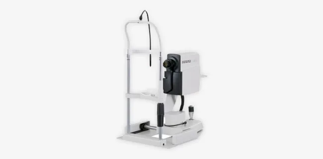 Angiography
HRA2 fluorescein angiography
University-hospital-grade angiography that examines the retina (fluorescein) and the choroid (indocyanine green) in a single session. Using an intravenous dye, it captures high-resolution fluorescein angiography and autofluorescence images of the retinal vessels.
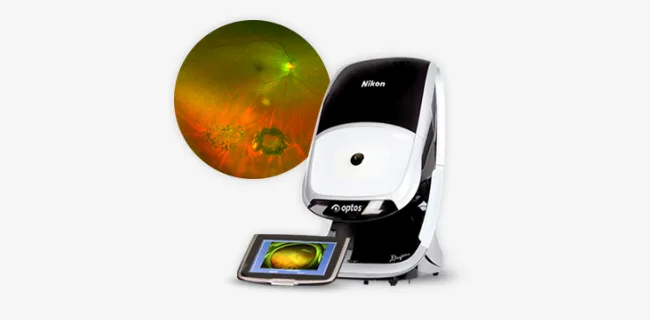 Wide-field imaging
Daytona (Optos) fundus camera
A high-resolution camera that photographs the retina at a wide angle without dilating drops. Screens for retinal disease and glaucoma, and checks for weakening or damage from the retina to the optic nerve.
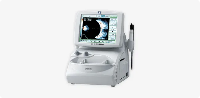 Ultrasound
B-Scan ocular ultrasound
Ultrasound imaging of the retina. When vitreous hemorrhage or opacity makes fundus photography impossible, B-Scan still allows diagnosis and differentiation of what is happening behind it.
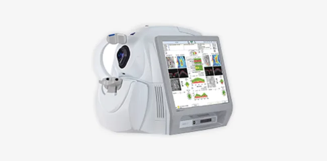 OCT
OCT
Cirrus HD-OCT
Optical coherence tomography that diagnoses whether visual field damage is progressing, and helps confirm glaucoma, retinal disease, optic nerve disease and brain-related problems.
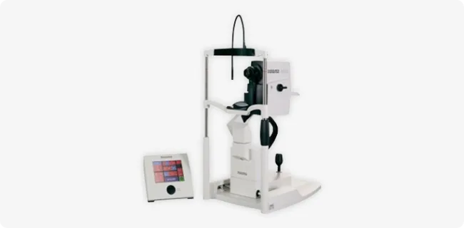 OCT · OCT-A
Spectralis OCT
Compares past and present scans to measure fine changes in a lesion and reads the state of the optic nerve fiber layer in detail. Its non-invasive OCT angiography examines retinal and choroidal vessels without dye.
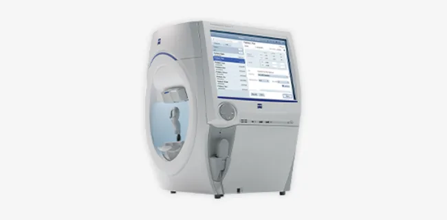 Visual field
HFA850 (Humphrey 850) field analyzer
Measures how far the visual field has narrowed and diagnoses whether the damage is progressing. Used to check glaucoma, retinal and optic nerve disease, and brain-related problems.
Which device answers which question?
| Device | Type | What it answers |
|---|---|---|
| HRA2 | Fluorescein + ICG angiography (IV dye) | Where retinal and choroidal vessels leak or grow abnormally |
| Daytona (Optos) | Non-mydriatic wide-field fundus camera | Wide-angle view of the retina without dilation; retina-to-optic-nerve screening |
| B-Scan | Ocular ultrasound | What lies behind a hemorrhage or opacity that blocks the view |
| Cirrus HD-OCT | Optical coherence tomography | Whether field damage is progressing; glaucoma, retina, optic nerve, brain |
| Spectralis OCT | OCT + non-invasive OCT-A | Fine lesion change over time; nerve fiber layer; vessels without dye |
| HFA850 | Visual field analyzer | How much field is lost and whether loss is advancing |
What operates when surgery is the answer?
When the diagnosis calls for surgery, treatment continues in-house on the retinal surgical suite - see how retinal treatments work.
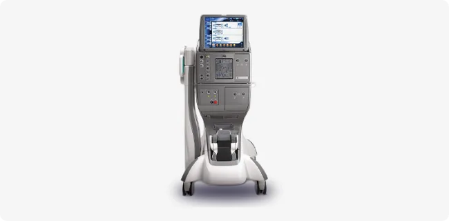 Vitrectomy platform
Alcon Constellation
A university-hospital-grade platform for micro-incision retinal surgery, cutting at up to 7,500 cycles per minute for controlled removal of the vitreous.
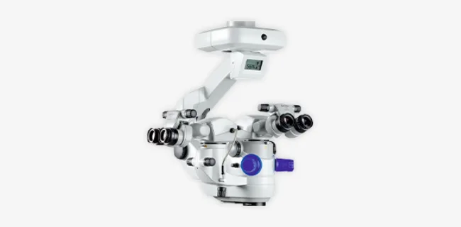 Surgical microscope
ZEISS Lumera 700
The operating microscope for retinal surgery, cataract surgery and corneal transplantation. Its clear operative view supports the accuracy of every step the surgeon takes.
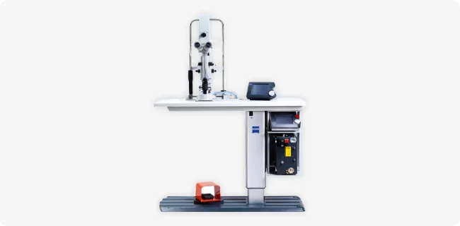 YAG laser
ZEISS VISULAS YAG III
A treatment laser used for acute angle-closure glaucoma and for after-cataract (posterior capsule opacification) following cataract surgery.
Questions about retinal examinations
Not necessarily. The Daytona (Optos) camera photographs the retina at a wide angle without dilating drops. Whether further dilated examination or angiography is needed depends on what the specialist finds on those first images.
That is what the B-Scan ultrasound is for. When vitreous hemorrhage or opacity makes fundus photography impossible, ultrasound still shows the state of the retina behind it, so the condition can be diagnosed and differentiated.
Often, yes. The Spectralis OCT includes non-invasive OCT angiography, which images retinal and choroidal vessels without dye. When a full dye study is needed, the HRA2 performs fluorescein and indocyanine green angiography through an intravenous injection.
Two ways. The Spectralis OCT compares past and present scans to measure microscopic change in a lesion, and the HFA850 visual field analyzer measures whether field loss is advancing. Together they show progression rather than a single snapshot.
Retina-trained specialists examine patients directly and, when surgery is needed, operate themselves. Meet the team on the Why GS Retina page.