Before an implantable collamer lens (ICL) is placed in the eye, the clinic has to answer two separate questions. The first is how strong the lens should be, which comes from your prescription. The second is how long the lens should be, so that it fits the space behind the iris without pressing on anything. That second answer cannot be read from your glasses. It comes from measurements of the inside of your eye: the depth of the space in front of the natural lens, the width of the eye, the health of the cell layer on the back of the cornea, and the eye pressure, retina and optic nerve. After surgery, the doctor checks the "vault", the small gap between the ICL and your natural lens, because that gap shows whether the chosen size is sitting as intended. Whether an ICL is suitable at all is decided by these exams, not by the prescription number.
This article explains what each pre-operative measurement is for, how lens size is usually chosen, what vault is and why it is measured more than once, the findings that can rule ICL out or ask you to wait, and how a trip to Seoul can be planned around the exam, the surgery and the follow-up.
Two numbers: lens power and lens size
Laser procedures such as SMILE and LASIK reshape the cornea, so the cornea's thickness and shape set their limits. An ICL works differently. It is a thin, flexible lens placed inside the eye, behind the coloured iris and in front of your own natural lens, and it corrects focus optically. The cornea is not reshaped. If you are still deciding between these routes, our comparison of SMILE, LASIK and ICL sets out how they differ.
Because the ICL is a physical object, it has two specifications that are worked out separately.
- Power. This is calculated from your prescription and other readings, such as corneal curvature, corneal thickness and the depth of the front chamber of the eye.
- Length (size). This is the overall length of the lens from one footplate to the other. It has to suit the space in which the lens rests, a narrow groove behind the iris called the ciliary sulcus. That space differs from eye to eye, even between people with the same prescription.
A lens of the right power but the wrong size can still cause problems. GS Eye Center's ICL page describes the two directions of error. A lens that is too large can obstruct the flow of fluid inside the eye, which can raise eye pressure and lead to glaucoma. A lens that is too small can move inside the eye, which can reduce the cells on the back of the cornea and may contribute to cataract. This is why measurement comes before lens choice.
The measurements taken before surgery
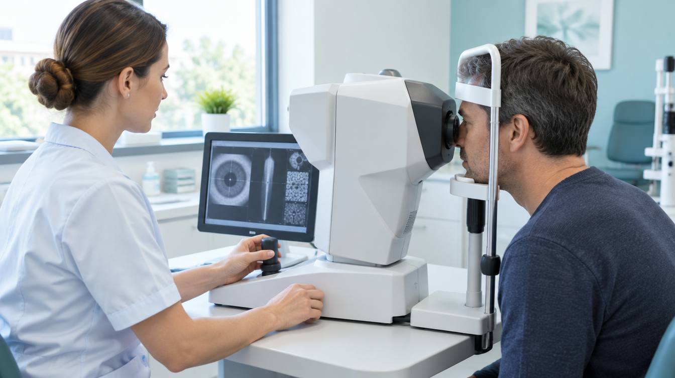
GS Eye Center lists five pre-operative exams for lens implantation: a vision and refraction exam, a lens position exam that measures the internal dimensions used for sizing and placement, an endothelial cell exam, an optic nerve exam for glaucoma risk, and a retina exam. The US Food and Drug Administration's patient information on phakic lenses describes a similar core, including pupil measurements, anterior chamber depth and endothelial cell counts.
| Measurement | What it is | Why it matters for ICL |
|---|---|---|
| Anterior chamber depth (ACD) | The distance from the inner surface of the cornea to the front of the natural lens | Shows whether there is room for a lens behind the iris. It is also one of the inputs for choosing lens size |
| Anterior chamber angle | The drainage angle where the iris meets the cornea, where fluid leaves the eye | A narrow angle raises concern about pressure problems once a lens is in place |
| Endothelial cell count | The number of cells per square millimetre in the single cell layer on the back of the cornea, which keeps the cornea clear | These cells do not grow back in meaningful numbers, so the count must be high enough for the years ahead. It also becomes a baseline for later checks |
| White-to-white distance | The horizontal width of the cornea, from the edge of the white on one side to the other | The most widely used input for estimating lens length, together with ACD |
| Sulcus-to-sulcus distance (in some clinics) | The width of the space behind the iris, measured with ultrasound | An alternative way to estimate lens length |
| Refraction, including with dilating drops | Your prescription, measured as you normally see and again with the focusing muscle relaxed | Sets the lens power. Prescription stability is checked against older records |
| Pupil size | How wide the pupil opens, including in dim light | Relevant to glare and halos at night |
| Eye pressure and optic nerve | Pressure readings and imaging or field testing of the optic nerve | Glaucoma or high pressure can rule ICL out or need treatment first |
| Retina | A dilated look at the back of the eye, often with wide-field imaging | High myopia is linked with retinal conditions that should be found and treated before surgery |
GS Eye Center's diagnostic equipment page lists instruments that record several of these readings, including anterior chamber depth and corneal diameter (GALILEI G4), anterior chamber depth and visible iris diameter (AL-SCAN optical biometer), endothelial cell counts (specular microscope), eye pressure and corneal thickness, retinal imaging (Spectralis and Cirrus OCT, OPTOS) and visual field testing (Humphrey). The page does not say which instrument's readings are used for ICL sizing in a given case. That is a reasonable question to ask at the consultation.
Contact lenses change the shape of the cornea slightly. The FDA advises that you may be asked to stop wearing them before the exam, anywhere from the day itself to a few weeks earlier, so the measurements describe your eye rather than the lens. Ask the clinic for the exact interval for the type of lens you wear.
How lens size is chosen
The most common method combines the horizontal white-to-white distance with the anterior chamber depth. The manufacturer of the EVO ICL, STAAR Surgical, provides sizing recommendations built on these two readings. In the original US clinical study of the Visian ICL, lens lengths from 12.1 to 13.7 mm were selected this way, and STAAR later adjusted its recommendations after analysing the study data.
Some surgeons also use ultrasound biomicroscopy to measure the sulcus-to-sulcus distance directly, because the lens rests in the sulcus rather than at the edge of the cornea. Anterior segment optical coherence tomography (OCT) is another way to image the front of the eye. A review of the published evidence by ophthalmologist Mark Packer found no clinically meaningful difference in the resulting vault between white-to-white-based and sulcus-to-sulcus-based sizing, and noted that white-to-white with ACD remains the most widely studied method.
The same review makes a point that matters for patients. Some of the variation in vault does not come from the sizing method. It comes from how the lens interacts with the individual anatomy behind the iris. In the studies reviewed, imaging-based sizing did not reduce this variation. Sizing is a careful prediction. The vault check after surgery shows how that prediction turned out.
GS Eye Center keeps four lens types: EVO+ ICL (STAAR Surgical, USA), KPL (Koryoeyetech, Korea) and GLAZE (Biotech, Switzerland), which sit behind the iris, and Artiflex (OPHTEC, Netherlands), which is fixed to the front of the iris. The vault discussion below applies to the three lenses that sit behind the iris. Which lens and which size suit your eye is decided by the surgeon after the exam.
What vault is, and why it is checked after surgery
Vault is the distance between the back surface of the ICL and the front surface of your natural lens. The ICL is designed to arch slightly over the natural lens instead of resting on it. That small space lets fluid move across the natural lens.
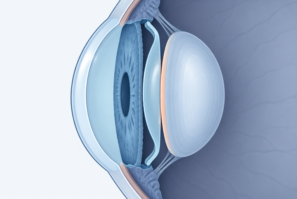
Published studies commonly describe a vault of about 250 to 750 micrometres (roughly a quarter to three quarters of a millimetre) as the target range. Outside that range, the concerns run in two directions.
- Low vault. When the ICL sits too close to the natural lens, contact between the two has been linked with clouding of the front of the natural lens (anterior subcapsular opacity) and cataract.
- High vault. When the ICL pushes too far forward, it can crowd the drainage angle, and has been associated with raised eye pressure, pupillary block and pigment loss from the iris.
The review mentioned above lists lens exchange or removal among the adverse events linked with vault that is too high or too low. GS Eye Center's Chinese-language ICL page names a lens that shifts position or sits at an unsatisfactory vault as one of the reasons a lens may need to be exchanged or removed.
Vault is not a fixed number. Studies have reported that vault becomes smaller when the pupil narrows, for example in bright light. It also changes over time. One study that measured vault at one week, one month, three months and six months after surgery found that it tended to decrease over that period, with the decrease slowing after about three months. That is why vault is measured at more than one visit, and why a reading on the first day is a starting point, not a final verdict.
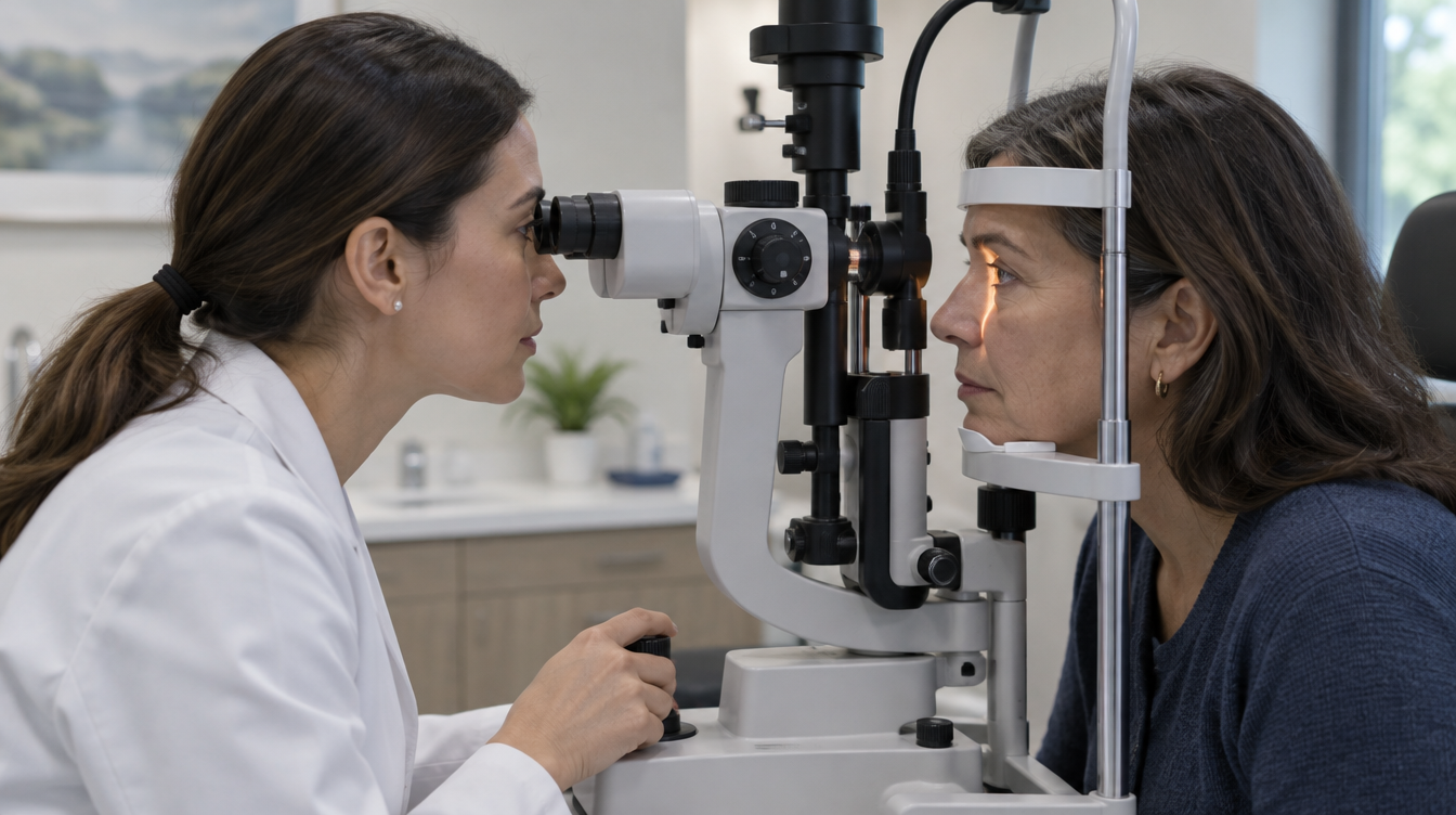
Vault is usually measured with a slit-lamp microscope, an imaging device such as anterior segment OCT, or both. Eye pressure is checked at the same visits, because a pressure rise and a high vault can be connected.
Findings that rule ICL out, or mean waiting
A suitable prescription is not enough on its own. GS Eye Center's Chinese-language ICL page lists situations in which ICL is not advised or another problem needs treating first: a shallow anterior chamber, a low endothelial cell count, glaucoma or a narrow drainage angle, cataract, inflammation inside the eye such as uveitis, untreated retinal disease, a prescription that is still changing, pregnancy or breastfeeding, and poorly controlled diabetes or autoimmune disease. The same page describes the general starting point as adults aged 18 or over with a stable prescription who are not suited to laser correction, with the decision based on chamber depth, endothelial cell count, eye length and retinal findings.
US labelling gives a useful sense of how strict the anatomy limits are. In the United States, the EVO ICL is indicated for people aged 21 to 60 (the upper limit was raised from 45 in February 2026) with an anterior chamber depth of at least 3.0 mm, measured from the inner surface of the cornea to the front of the natural lens. It is not used when the drainage angle is too narrow or the endothelial cell count is below an age-based minimum. Approved ranges differ by country. GS Eye Center's page gives 18 or over as the general starting age, and suitability is decided from your exam results.
The endothelial minimum depends on age for a simple reason. Endothelial cells decline slowly throughout life, and a younger person has more years ahead. In the US clinical study of the EVO lens, average cell density was 3.0% lower one year after surgery and 6.7% lower at three years than before surgery. The minimum counts in the US labelling are set by age and chamber depth so that, at the rates of loss seen in that study, the eye should still have at least 1,000 cells per square millimetre at age 75. A deeper chamber allows a lower minimum, and a younger age requires a higher one.
If laser correction was already ruled out for you and you are wondering what an ICL consultation involves, our article on what happens when laser is not the answer covers that conversation.
After surgery: check-ups and records to keep
The schedule below describes what is typical. Your surgeon sets the exact plan for your eyes and the lens used.
| When | What is usually checked | Why |
|---|---|---|
| A few hours after surgery | Eye pressure and lens position | Pressure can rise early, and the first vault reading is taken |
| Next day | Vision, eye pressure, vault, signs of inflammation | Confirms the lens is centred and the eye is settling |
| First weeks | Vault, pressure, healing of the incision, drop schedule | Vault tends to change most in the first months |
| First months | Vault, pressure, endothelial cell count, refraction | Shows how vault and vision have settled |
| Every year, for life | Endothelial cell count, eye pressure and a full eye exam | The lens is a long-term implant, and changes can develop slowly without symptoms |
The FDA's patient information says that people with a phakic lens need regular follow-up with an eye doctor for the rest of their lives, with endothelial cell counts repeated over time and the results kept so that the rate of change can be estimated. Keep the implant identification card you receive, and show it to any eye doctor who treats you later, especially before cataract surgery or any other procedure inside the eye.
GS Eye Center's aftercare advice for ICL includes using drops as prescribed and having eye pressure measured on schedule, not rubbing or pressing on the eye, and avoiding swimming, saunas and strenuous exercise in the early period. Seek care promptly, and contact the clinic, if you notice a sudden drop in vision, an aching eye, headache or light sensitivity. The FDA also advises seeing an eye doctor right away for flashes of light, floating spots or blank spots in your vision, which can be signs of a retinal problem, and wearing protective eyewear for sports or work that could injure the eye.
Planning the exam, surgery and follow-up from North America
For international patients, the exam and surgery usually happen on the same trip. The exam confirms whether ICL is suitable and which lens fits, the surgeon confirms the procedure, and surgery follows within your travel window.
- Send records first. Your glasses and contact lens prescriptions over the past few years and any previous eye scans help the team judge whether the trip makes sense. They do not replace the exam, because chamber depth, endothelial cell count and the internal measurements for sizing are not in those records.
- Allow for the exam day. GS Eye Center's exam visit takes about two hours. If dilating drops are needed, add about 30 minutes, and do not plan to drive afterward.
- Ask how many days separate the exam and the surgery. The lens type, size and power are chosen from your measurements. Your coordinator plans those days with you before you book flights.
- Plan for at least the early checks. Vault and eye pressure are usually checked within hours and again the next day. How long you stay after that is set case by case at the consultation.
- Arrange care at home. Find an eye doctor near home who can measure eye pressure and endothelial cell count and can assess the lens position. Take copies of your pre-operative measurements, the lens details and the first vault readings.
- Contact lens break. Ask how long to stop wearing contact lenses before the exam, and do not wear them on the flight.
GS Eye Center (GS안과) is an eye clinic at Gangnam Station Exit 1, Seoul, open since 2005. It performs implantable lens surgery with EVO+ ICL, KPL, GLAZE and Artiflex lenses, and the lens type and size for each patient are decided after the pre-operative exams.
This article was written from GS Eye Center's published information and the doctors' explanations. It is general information, not a diagnosis or a treatment recommendation for any individual.