Corneal cross-linking (CXL) is a treatment that makes a weakening cornea stiffer. Riboflavin (vitamin B2) drops are soaked into the cornea and then activated with ultraviolet-A light, which helps form new bonds between the cornea's collagen fibres. It is used mainly for keratoconus and for corneal bulging (ectasia) after laser eye surgery, when scans show the cornea is changing shape. The aim is to slow or stop that change. Cross-linking does not undo thinning or bulging that has already happened, and it is not a way to stop wearing glasses. Most people still need glasses or contact lenses afterward, often with a new prescription once the eye settles. Whether cross-linking is needed, and in which form, is decided after corneal scans and an exam.
This article covers what keratoconus and ectasia are, what cross-linking does and does not do, the difference between the main methods in plain terms, how the screening exam picks up a cornea at risk, and what the first week and month of recovery usually look like. It also explains why a scan sometimes leads to the answer that LASIK or SMILE is not suitable, and how a visit to Seoul can be planned around follow-up exams.
Keratoconus and ectasia: when the cornea starts to change shape
The cornea is the clear dome at the front of the eye. It does most of the eye's focusing, so its shape matters. In keratoconus, the cornea gradually thins and bulges forward until it is shaped more like a cone. Light no longer focuses cleanly, and vision becomes blurry and distorted.
Keratoconus often starts in the late teens or early twenties. In many people it worsens slowly over 10 to 20 years, but in some it moves faster. Early signs can include:
- mild blurring, or straight lines that look slightly bent or wavy
- more sensitivity to light and glare
- astigmatism or nearsightedness that keeps increasing, so glasses need changing often
- contact lenses that become hard to wear
Doctors do not know exactly why it develops. About one in ten people with keratoconus has a parent with it. It is also linked with eye allergies, heavy eye rubbing and some connective tissue disorders. Rubbing matters enough that eye specialists put "stop rubbing your eyes" near the top of their advice, and treating itchy allergies is part of that.
Ectasia is the general name for a cornea that thins and bulges. Keratoconus is the most common form. Rarely, the same kind of bulging appears after LASIK or other laser vision correction, because removing tissue has weakened a cornea that was already less sturdy than it looked.
Glasses, soft lenses and later rigid or specialty contact lenses help people see more clearly, and ring implants or a corneal transplant are options in advanced cases. Cross-linking has a different job. It does not sharpen the focus. It targets the weakening itself.
What cross-linking does, and what it does not do
Collagen fibres give the cornea its strength, a little like the threads in woven fabric. In keratoconus the links holding those fibres together are fewer or weaker, so the tissue can stretch. During cross-linking, riboflavin absorbs ultraviolet-A light and sets off a chemical reaction that forms new bonds between neighbouring collagen molecules. Oxygen in the tissue is part of that reaction. The effect is strongest in the front part of the cornea, where the riboflavin concentration is highest.
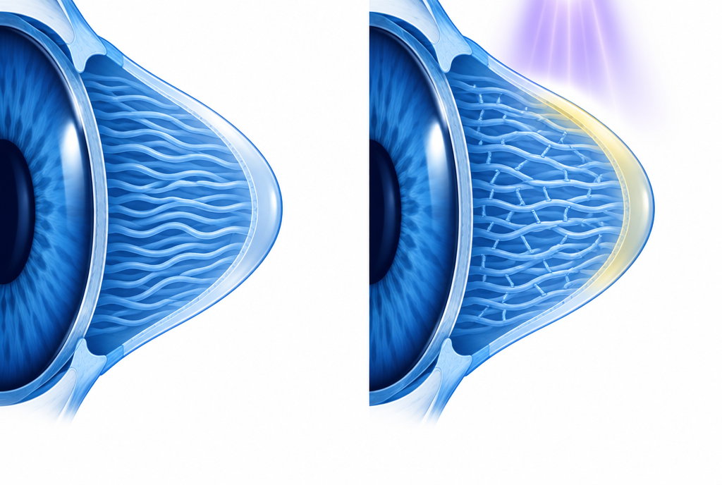
The goal is a stiffer cornea that holds its shape. In the United States, the FDA approved cross-linking with riboflavin and ultraviolet-A light in 2016 for progressive keratoconus and for corneal ectasia after refractive surgery, based on year-long studies.
It helps to be clear about what to expect:
- It aims to stop the change, not reverse it. An existing cone or bulge does not return to normal.
- It is not vision correction. Glasses or contact lenses are usually still needed. If sharpness improves a little, treat it as a possible extra, not the purpose.
- Vision is worse before it settles. After the standard method, vision is very blurry at first and improves over the following weeks. The glasses or contact lens prescription often changes for several months.
- It may avoid bigger surgery for some people. By keeping keratoconus from advancing, cross-linking can mean a corneal transplant is never needed. That is a hope for some eyes, not a promise for every eye.
Epithelium-off, epithelium-on, standard and accelerated
The cornea's outer skin, the epithelium, is a barrier that riboflavin does not cross easily. The main methods differ in how they deal with that barrier and in how the light is delivered.
Epithelium-off. This is the standard approach and the one with the most research behind it. After numbing drops, the central epithelium is gently removed so riboflavin can reach the tissue beneath. In the original protocol, riboflavin drops are applied for about 30 minutes and the light is then applied for close to 30 minutes. A soft bandage contact lens goes on afterward, usually for about a week, while the surface grows back. The surface layer typically closes over within three to four days.
Epithelium-on (transepithelial). The outer skin is left in place, and the riboflavin is formulated or delivered so that it can pass through. This tends to mean less discomfort and a quicker recovery. Some reviews describe it as less consistent at halting progression, and a 2021 Cochrane review found it is still unknown whether either approach has an advantage. In October 2025 the FDA approved an epithelium-on cross-linking system for keratoconus in people aged 13 and older.
Standard versus accelerated. Accelerated protocols deliver a similar amount of light energy at a higher intensity over a shorter time, so the light step takes minutes rather than about half an hour. Many eye specialists now use accelerated settings, and studies continue on how to balance time and intensity.
Cross-linking added to laser vision correction. Here the purpose is preventive rather than treating established disease. Right after SMILE, LASIK or LASEK, riboflavin is applied to the treated tissue, rinsed off, and a short light treatment follows. GS Eye Center describes this add-on, sometimes called "LASIK Xtra", as taking about three to five minutes on its Avedro KXL systems. Published evidence on this preventive use is more limited than for keratoconus treatment, so it is discussed case by case after the exam.
How the screening exam spots a cornea at risk
Moderate or advanced keratoconus can be seen with a microscope. Early keratoconus often cannot, and that is where corneal imaging matters.
- Topography maps the curvature of the front surface of the cornea.
- Tomography maps both the front and back surfaces and the thickness at every point. Early changes often appear on the back surface or in the thickness pattern before they show up in the front curvature or in your glasses prescription.
International keratoconus experts list abnormal elevation of the back surface, an abnormal thickness pattern and corneal thinning as key findings, and they recommend tomography as a standard part of screening before laser vision correction. At GS Eye Center the corneal workup includes Pentacam and GALILEI G4 tomography, K5M-TOPO topography and corneal thickness measurement, as part of an exam visit that takes about two hours. Our guide to what the screening exam measures explains each test.
Progression needs two points in time. A single scan shows the shape of the cornea today. Deciding that keratoconus is getting worse means comparing scans taken months apart, looking for steepening of the front or back surface or further thinning. That is why earlier scans and old prescriptions from home are worth bringing. In children and teenagers, keratoconus can move quickly, so doctors may not wait as long for proof of change.
Reasons to wait or choose differently. GS Eye Center's Chinese-language cross-linking page lists situations in which cross-linking is not advised or something else needs treating first: a cornea thinner than the minimum the treatment requires, corneal scarring or poor surface healing, a history of herpes infection of the cornea, active eye infection or inflammation, severe dry eye, autoimmune disease, and pregnancy or breastfeeding.
Why some people are told LASIK or SMILE is not suitable
Laser vision correction works by removing a small amount of corneal tissue. In a healthy cornea that is well tolerated. In a cornea already inclined to bulge, removing tissue can weaken it further and raise the risk of ectasia. For that reason, the latest international consensus on keratoconus states that LASIK is generally contraindicated in keratoconus and in eyes prone to ectasia. Surgeons apply similar caution to other procedures that remove corneal tissue, such as SMILE.
There are three common situations behind a "no":
- Keratoconus is confirmed. The first goal becomes keeping the cornea stable. Vision is then usually managed with glasses or contact lenses, and other treatments are considered separately.
- The scan is suspicious but not conclusive. The doctor may advise against surgery for now, or ask for a repeat scan later to see whether anything is changing.
- The cornea is thin for the prescription. Thin is not the same as weak. A thin cornea with a normal shape may still have options, which is a separate question from keratoconus.
GS Eye Center's cross-linking page places the treatment in these planning decisions in two ways. It is used to treat keratoconus itself. It can also be added at the same session as SMILE, LASIK or LASEK for eyes the surgeon judges to be at higher risk, such as a family history of keratoconus, thin corneas or farsightedness, high myopia that needs a deeper treatment, long-term contact lens wear, or concern about the prescription creeping back. It is not combined with ICL, because a lens implant does not reshape the cornea. The page also notes that some people who were turned down for laser surgery because keratoconus was suspected may be re-evaluated after cross-linking. That is an individual medical judgment made from the scans, not a standard route to laser surgery.
If you have already been told no elsewhere, our article on being turned down before explains what a second look can and cannot change.
Recovery: the first week, the first month and after
The timeline below describes standard epithelium-off cross-linking as a treatment on its own. When cross-linking is added to SMILE, LASIK or LASEK, recovery and follow-up visits follow the plan for that main procedure.
| Stage | What is common | When to contact the clinic |
|---|---|---|
| Treatment day | Numbing drops during the procedure; afterward, blurry vision, watering and a bandage contact lens on the eye | Severe pain that the prescribed medicine does not ease |
| First few days | A gritty or something-in-the-eye feeling, light sensitivity, discomfort or mild pain while the surface grows back, often over three to four days | Sudden drop in vision, marked pain, or more redness or discharge |
| Around one week | Bandage lens removed at a check-up; still no rubbing, no water in the eye, no eye makeup, no heavy exercise, no smoky or dusty places during the first week | Vision getting worse instead of slowly better |
| Weeks two to three | Vision that started very blurry gradually improves; most people are back to normal daily activities within one to two weeks | Pain or light sensitivity that returns after it had eased |
| Weeks six to eight | Prescription still shifting; glasses or rigid contact lenses are often refitted around this time | Any sudden change in vision |
| Following months | Vision may not be fully stable for several months; temporary haze is common and usually fades; repeat scans check that the cornea has stopped changing | New blurring or glare between scheduled checks |
Antibiotic and steroid drops are usually prescribed to help the eye heal. Follow the drop schedule exactly. GS Eye Center also advises wearing sunglasses outdoors with good UV protection and avoiding swimming, saunas and strenuous exercise in the early period.
Rarer problems include infection, scarring, slow healing of the surface, lasting haze and, in a small number of eyes, keratoconus that keeps worsening despite treatment. This is why the follow-up scans matter, even when the eye feels comfortable.
Planning cross-linking around a trip to Seoul
The practical plan depends on which form of cross-linking you have, and that is decided after the exam.
- Send your records first. Recent scans, your glasses and contact lens prescriptions over the past few years and any diagnosis letters help the team see whether the cornea has been changing. The final plan is made after the in-person exam.
- Cross-linking on its own. With the epithelium-off method, the bandage lens is usually removed about a week after treatment, so plan to stay near the clinic through that check. The clinic sets the exact schedule.
- Cross-linking added to SMILE, LASIK or LASEK. The add-on takes a few minutes in the same session, and your stay is planned around the main procedure.
- Flying. Cabin air is dry, so keep your drops in your hand luggage. Fly only when the doctor has cleared you.
- Back home. Find an eye doctor near home who can do corneal topography or tomography, and take copies of your scans from before and after treatment. Comparing scans over months and years is how stability is confirmed. A new glasses or contact lens fitting often happens at home around six to eight weeks.
- Keep the habits. Not rubbing your eyes and treating itchy allergies remain important after cross-linking.
GS Eye Center (GS안과) is an eye clinic at Gangnam Station Exit 1, Seoul, open since 2005. It performs cross-linking on four Avedro KXL systems with VibeX riboflavin, both as a treatment for keratoconus and as an add-on to SMILE, LASIK or LASEK, and the technique for each patient is discussed after the examination.
This article was written from GS Eye Center's published information and the doctors' explanations. It is general information, not a diagnosis or a treatment recommendation for any individual.